Veterinary CT
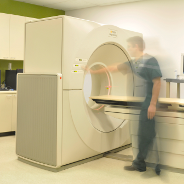
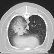
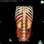
Computed Tomography (CT or CAT) is a noninvasive diagnostic imaging procedure that uses a combination of X-ray and computer technology to produce cross-sectional images (often called slices) of the body, making the images much more detailed than X-rays or even ultrasound. A CT scan shows highly detailed images of any part of the body but is best for the bones, nose, ears, GI, and lungs. Contrast CT is useful for assessing the urinary tract. CT studies also have the added advantage of speed—the entire scan can often be completed in minutes. Furthermore, the images can easily be used to create 2-D and 3-D reconstructions.
CT is particularly effective in evaluating:
- Chest tumors and lung masses
- Bone evaluation, particularly complex fractures
- Nasal, sinus and inner ear polyps, tumors or other masses
- Specific spinal conditions, such as calcified disc disease
- Ectopic ureter, when contrast is used
- Gastrointestinal evaluation
- Metastatic disease to the lungs
- Surgical planning
CT can also be used for:
- Soft tissue evaluation, when contrast is used
- Brain imaging (pituitary gland)
- Tumor evaluation, particularly for metastatic disease
If you are trying to determine if CT or MRI is most appropriate for your needs, just give us a call. One of our veterinary radiologists can answer any questions you may have.